

By Michelle Jaffee
A new high-tech imaging study led by neuroscientists at the University of Florida and collaborators in South Carolina and Istanbul, Turkey, adds to the burgeoning understanding of the brain’s waste-removal system, illuminating the role of key lymphatic structures whose disruption might contribute to pathology in neurodegenerative diseases, traumatic brain injury and multiple sclerosis.
The study, published in the journal Nature Communications, was led by UF neuroradiologist Mehmet Sait Albayram, M.D., and neurobiologist Onder Albayram, Ph.D., of the Medical University of South Carolina.
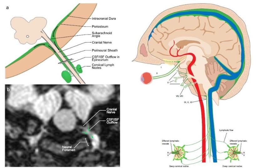
The retrospective study of 81 human participants focused on meningeal lymphatic vessels, analyzing 3D volumetric MRI images to trace naturally produced protein-rich lymphatic fluid, rather than using artificial contrast media. The investigators newly described the major human dural lymphatic structures along the dural venous sinuses in dorsal regions of the brain and along cranial nerves in the ventral regions.
Until recently, said Mehmet Sait Albayram, the processes by which the brain clears cellular waste have largely remained a mystery. Recent animal studies led to the discovery of lymphatic vessels around brain tissue and along blood vessels supplying the brain and also have shown how waste is drained toward lymph nodes in the neck, he said. Moreover, human imaging and autopsy studies have yielded evidence of similar brain lymphatics in humans as the knowledge base continues to expand.
“These findings provide a more complete picture than previous studies of the systems used for waste removal from the brain,” he said. “We hope that this and similar techniques may be used to advance our understanding of the basic physiology of human brain lymphatics, such as changes that might occur during sleep or stress.”
The study also newly revealed evidence of direct connections between lymphatic fluid channels along the cranial nerves and vascular structures to cervical lymph nodes. In addition, the research team identified age-related cervical lymph node atrophy and thickening of lymphatics channels in the dorsal and ventral regions.
“We believe this approach may provide insights on changes in brain lymphatic function that may occur in neurological conditions such as traumatic brain injury, Alzheimer’s disease and multiple sclerosis,” Mehmet Sait Albayram said.